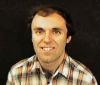
Paul Maccabee (1944–2023)

 |
|
| Paul Maccabee (1944–2023). Photo used with permission from the Downstate Health Sciences University website. |
My friend and collaborator Paul Maccabee died on April 24. Paul was a pioneer in the field of magnetic stimulation, a topic that Russ Hobbie and I discuss in Chapter 8 of Intermediate Physics for Medicine and Biology. Paul’s career and mine had many parallels. We both worked on magnetic stimulation in the late 1980s and early 1990s. We both collaborated with a leading neurophysiologist: Paul with Vahe Ammasian and me with Mark Hallett. We both recognized the importance of laboratory animal experiments for identifying physiological mechanisms. We both were comfortable working with biomedical engineers, I entered that field from physics and Paul from medicine.
Paul was about 15 years older than me and I viewed him as a role model. I believe I first met him at the 1989 International Motor Evoked Potential Symposium in Chicago, a key early conference dedicated to magnetic stimulation. Our paths crossed at other scientific meetings and his research had a major impact on my own. For years I taught a graduate class on bioelectricity at Oakland University and I had my students read Paul’s 1993 Journal of Physiology paper (described below) which I assigned because it’s a classic example of a well-written scientific article. According to Google Scholar that paper has been cited 374 times, and it should be cited even more.
I wrote about Paul in my review of the development of transcranial magnetic stimulation (BOHR International Journal of Neurology and Neuroscience, Volume 1, Pages 8–20, 2022, https://doi.org/10.54646/bijnn.002). Below I quote part of that article. I put his name in bold so you can find it easily.
Although this experiment [performed by Jan Nilsson and Marcela Panizza at the National Institutes of Health, see reference 49] confirmed [Peter Basser and my] prediction [that neural excitation occurs where the gradient of the induced electric field is largest, see reference 58], there were nevertheless concerns because of the heterogeneous nature of the bones and muscles in the human arm. At about the same time Nilsson and Panizza were doing their experiment at NIH, Paul Maccabee was performing an even better experiment at the New York Downstate Medical Center in Brooklyn. Maccabee obtained his MD from Boston University and collaborated in Brooklyn with the internationally acclaimed neuroscientist Vahe Ammasian [1, 40–43]. This research culminated in their 1993 article in the Journal of Physiology, in which they examined magnetic stimulation of a peripheral nerve lying in a saline bath [44]. First, they measured the electric field Ey (they assumed the nerve would lie above the coil along the y-axis) and its derivative along the nerve produced by a figure-8 coil located under the bath (Figure 9). They found that the electric field was maximum directly under the center of the coil, but the magnitude of the gradient dEy/dy was maximum a couple centimeters either side of the center.
 |
Figure 9. Contour plots of the electric field (Ey, red) and its spatial derivative (dEy/dy, blue) induced by a figure-eight coil (purple) placed under a tank filled with saline and measured using a bipolar recording electrode. The y direction is downward in the figure, parallel to the direction of the nerve (see Figure 10). Adapted from Figure 2 of Maccabee et al. [44]. |
Next they placed a bullfrog sciatic nerve in the dish and recorded the electrical response from one end (Figure 10). They found a 0.9 ms delay between the recorded action potentials when the polarity of a magnetic stimulus was reversed (the yellow and red traces on the right). Given a propagation speed of about 40 m/s, the shift in excitation position was about 3.6 cm, consistent with what Basser and I would predict.
 |
Figure 10. Recordings from an electrode (black dot) at the distal end of a bullfrog sciatic nerve (green) that was immersed in a trough filled with saline (blue) and stimulated with a figure-8 coil (purple). The nerve emerged from the saline to rest on the recording electrode in air. The compound nerve action potentials were elicited by a stimulus of one polarity (orange), then the other (red). Adapted from Figure 3 of Maccabee et al. [44]. |
So far, their study was similar to what we performed at NIH in a human, but then they did an experiment that we could not do. To determine how a heterogeneity would impact their results, they placed two insulating cylinders on either side of the nerve (Figure 11). These cylinders modified the electric field, moving the negative and positive peaks of the activating function closer together. They observed a corresponding reduction in the latency shift. This experiment provided insight into what happens when a human nerve passes between two bones, or some similar heterogeneity.
Figure 11. Magnetic stimulation of a sheep phrenic nerve immersed in a homogeneous (left) and inhomogeneous (right) volume conductor. The figure-8 coil (purple) was positioned under the nerve (green). The yellow circles indicate the position of the insulating cylinders. The electric field Ex (red) and its gradient dEx/dx (blue) were measured along the nerve trajectory. The compound nerve action potentials at the recording electrode were measured for a magnetic stimulus of one polarity (orange) and then the other (green). Adapted from Figure 5 of Maccabee et al. [44]. Finally, they changed the experiment by bending the nerve and found that a bend caused a low threshold “hot spot,” and that excitation at that spot occurred where the electric field, not its gradient, was large. This result was consistent with Nagarajan and Durand’s analysis of excitation of truncated nerves [47].

In my opinion, Maccabee’s [44] article is the most impressive publication in the magnetic stimulation literature. Only Barker’s original demonstration of transcranial magnetic stimulation can compete with it [2].
Later in that review, I discussed a collaborative paper that Paul and I published.
One frustrating feature of the activating function approach is that excitation does not occur directly under the center of a figure-8 coil, where the electric field is largest, but off to one side, where the gradient peaks (Figure 9). Medical doctors do not want to guess how far from the coil center excitation occurs; they would prefer a coil for which “x” marks the spot. It occurred to me that such a coil could be designed using two adjacent figure-8 coils. I called this the four-leaf coil (Figure 12). John Cadwell from Cadwell Laboratories (Kennewick, Washington) built such a coil for me. Having seen the excellent results that Maccabee was obtaining using his nerve-in-a-dish setup, I sent the coil to him so he could test it. The resulting paper [65] showed that for one polarity of the stimulus the magnitude of the gradient of the electric field was largest directly under the coil center so the axons there were depolarized (“x” really did mark the spot of excitation). In addition, if the polarity of the stimulus was reversed, the magnitude of the gradient remained large under the coil center, but it now tended to hyperpolarize rather than depolarize the axons. Maccabee and I hoped that such hyperpolarization could be used to block action potential propagation, acting like an anesthetic. The Brooklyn experiments verified all the predictions of the activating function model for the four-leaf coil. Unfortunately, Maccabee never observed any action potential block. Perhaps, the hyperpolarization required for block was greater than the coil could produce.
Figure 12. A four-leaf coil (purple) used to stimulate a peripheral nerve (blue). Adapted from Figure 1 of Roth et al. [65]. [1] Amassian, V. E., Eberle, L., Maccabee, P. J., and Cracco, R. Q. (1992). Modelling magnetic coil excitation of human cerebral cortex witha peripheral nerve immersed in a brain-shaped volume conductor:The significance of fiber bending in excitation. Electroenceph. Clin. Neurophysiol., 85, 291–301.
[2] Barker, A. T., Jalinous, R., and Freeston, I. L. (1985). Non-invasive magnetic stimulation of human motor cortex. Lancet, 1, 1106–1107.
[40] Maccabee, P. J., Amassian, V. E., Cracco, R. Q., and Cadwell, J. A. (1988). An analysis of peripheral motor nerve stimulation in humans using the magnetic coil. Electroenceph. Clin. Neurophysiol., 70, 524–533.
[41] Maccabee, P. J., Eberle, L., Amassian, V. E., Cracco, R. Q., Rudell, A., and Jayachandra, M. (1990). Spatial distribution of the electric field induced in volume by round and figure ‘8’ magnetic coils: Relevance to activation of sensory nerve fibers. Electroenceph. Clin. Neurophysiol., 76, 131–141.
[42] Maccabee, P. J., Amassian, V. E., Cracco, R. Q., Cracco, J. B., Eberle, L., and Rudell, A. (1991a). Stimulation of the human nervous system using the magnetic coil. J. Clin. Neurophysiol., 8, 38–55.
[43] Maccabee, P. J., Amassian, V. E., Eberle, L. P., Rudell, A. P., Cracco, R. Q., Lai, K. S., and Somasundarum, M. (1991b). Measurement of the electric field induced into inhomogeneous volume conductors by magnetic coils: Application to human spinal neurogeometry. Electroenceph. Clin. Neurophysiol., 81, 224–237.
[44] Maccabee, P. J., Amassian, V. E., Eberle, L. P., and Cracco, R. Q. (1993). Magnetic coil stimulation of straight and bent amphibian and mammalian peripheral nerve in vitro: Locus of excitation. J. Physiol., 460, 201–219.
[47] Nagarajan, S. S., Durand, D. M., and Warman, E. N. (1993). Effects of induced electric fields on finite neuronal structures: A simulation study. IEEE Trans. Biomed. Eng., 40, 1175–1188.
[49] Nilsson, J., Panizza, M., Roth, B. J., Basser, P. J., Cohen, L. G., Caruso, G., and Hallett, M. (1992). Determining the site of stimulation during magnetic stimulation of a peripheral nerve. Electroenceph. Clin. Neurophysiol., 85, 253–264.
[58] Roth, B. J. and Basser, P. J. (1990). A model of the stimulation of a nerve fiber by electromagnetic induction. IEEE Trans. Biomed. Eng., 37, 588–597.
[65] Roth, B. J., Maccabee, P. J., Eberle, L., Amassian, V. E., Hallett, M., Cadwell, J., Anselmi, G. D., and Tatarian, G. T. (1994a). In-vitro evaluation of a four-leaf coil design for magnetic stimulation of peripheral nerve. Electroenceph. Clin. Neurophysiol., 93, 68–74.

Although my name was listed first on our joint 1994 article, Paul could easily have been the lead author. The coil shape was my idea but he performed all the experiments. I never set foot in Brooklyn; I just mailed the coil to him.
Paul was a giant in the field of magnetic stimulation. The articles I list above are only a few of the many he published. For a medical doctor he had a strong grasp of electricity and magnetism. I lost track of him over the years but had the good fortune to reconnect with him a few months ago by email.
I miss Paul Maccabee. Anyone who studies, uses, or benefits from magnetic stimulation owes him a debt of gratitude. I know I do.
Source: http://hobbieroth.blogspot.com/2023/08/paul-maccabee-19442023.html
Anyone can join.
Anyone can contribute.
Anyone can become informed about their world.
"United We Stand" Click Here To Create Your Personal Citizen Journalist Account Today, Be Sure To Invite Your Friends.
Please Help Support BeforeitsNews by trying our Natural Health Products below!
Order by Phone at 888-809-8385 or online at https://mitocopper.com M - F 9am to 5pm EST
Order by Phone at 866-388-7003 or online at https://www.herbanomic.com M - F 9am to 5pm EST
Order by Phone at 866-388-7003 or online at https://www.herbanomics.com M - F 9am to 5pm EST
Humic & Fulvic Trace Minerals Complex - Nature's most important supplement! Vivid Dreams again!
HNEX HydroNano EXtracellular Water - Improve immune system health and reduce inflammation.
Ultimate Clinical Potency Curcumin - Natural pain relief, reduce inflammation and so much more.
MitoCopper - Bioavailable Copper destroys pathogens and gives you more energy. (See Blood Video)
Oxy Powder - Natural Colon Cleanser! Cleans out toxic buildup with oxygen!
Nascent Iodine - Promotes detoxification, mental focus and thyroid health.
Smart Meter Cover - Reduces Smart Meter radiation by 96%! (See Video).


| Online: | |
| Visits: | 1,601,827,659 |
| Stories: | 8,145,599 |
Whistler Blowers, Insiders
